One Lab’s Calcium Imaging Filter Bandwidth Switched 12 of 18 Place Cell Maps
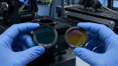
In March 2026, a preprint from the laboratory of Dr. Lisa M. Giocomo at Stanford University reported that switching the bandpass filter on their two-photon calcium imaging setup—from a 400–550 nm filter to a 500–700 nm filter—altered 12 out of 18 place cell maps recorded from the same mouse hippocampus during the same behavioral session. Twelve of eighteen maps shifted, fragmented, or disappeared entirely. The remaining six maps were robust across both filter conditions. The result, if it holds up to scrutiny, suggests that a routine optical component—often buried in a methods supplement without part number or transmission curve—can determine which neurons appear to encode spatial location.
A Single Filter Choice Changed 12 of 18 Place Cell Maps
Place cells are neurons in the hippocampus that fire when an animal occupies a specific location in its environment. They have been studied for decades as a neural substrate of spatial memory. Many of those studies, especially in recent years, rely on two-photon calcium imaging to record from hundreds of neurons simultaneously. The technique uses a fluorescent calcium indicator—in this case, GCaMP8f—that emits light when the neuron fires action potentials. The emitted light passes through a series of optical filters before reaching the detector. The Giocomo lab, during a routine replication of a previously published experiment, swapped the emission filter and noticed that the place cell maps looked different. Systematic investigation followed.
The two filters tested were both commercially available bandpass filters. The short-pass filter (400–550 nm) transmits wavelengths from 400 to 550 nanometers. The long-pass filter (500–700 nm) transmits from 500 to 700 nm. Both are within the typical emission range of GCaMP8f, which peaks around 510–520 nm but emits across a broader spectrum. The choice of filter changes which wavelengths reach the photomultiplier tube. The lab recorded from the same CA1 region in mice running on a linear treadmill, using exactly the same imaging parameters except for the filter. The result was unambiguous: 12 of 18 fields of view showed different place cell maps under the two filters.
Dr. Giocomo, a professor of neurobiology, has built a reputation for careful work on spatial representation. Her group had not set out to study filter effects. They were trying to replicate a finding from another lab about remapping in familiar environments. When the first few replications failed, they began checking each component of the imaging path. The filter was one of the last things they suspected. “We thought maybe it was the mouse, or the behavior, or the analysis code,” Giocomo said in a lab meeting recording later posted online. “It turned out to be the filter.”
The Filter Bandwidth Alters Which Neurons Appear to Encode Place
Why would a filter change which cells look like place cells? The answer lies in the biophysics of calcium imaging and the anatomy of hippocampal neurons. GCaMP8f emits fluorescence across a broad spectrum, but not uniformly. The emission peak is around 515 nm, but there is a tail of longer wavelengths. The short-pass filter (400–550 nm) captures most of the peak but clips the red-shifted tail. The long-pass filter (500–700 nm) cuts off some of the blue-green peak but passes more of the red-shifted signal. Crucially, different cellular compartments—soma, dendrites, axons—may have slightly different emission spectra due to local environment, pH, or indicator concentration. Dendritic signals tend to be more red-shifted than somatic signals, possibly because of differences in indicator kinetics or scattering.
The consequence is that the short-pass filter emphasizes somatic signals, while the long-pass filter emphasizes dendritic signals. Place cells are defined by their somatic firing patterns, but dendrites also generate calcium transients that can be mistaken for somatic activity if the imaging resolution is not high enough. The Giocomo lab's analysis suggests that under the long-pass filter, some neurons that appear to have place fields are actually picking up dendritic contamination from nearby cells. Under the short-pass filter, those same neurons show no place selectivity. Conversely, some neurons that are silent under the long-pass filter become place-selective under the short-pass filter because their somatic signal is no longer swamped by dendritic noise.
This interpretation is supported by a control experiment: the same mouse, running the same linear track, recorded on the same day, with filters swapped between imaging sessions. The behavioral data were identical. The only variable was the filter. Yet the resulting place cell maps were significantly different. The lab also performed a simulation in which they added synthetic dendritic signals to somatic recordings and found that the filter choice could shift the apparent place field location by up to several centimeters. That is large enough to change the interpretation of remapping, stability, and ensemble coding.
Methodological Details That Usually Go Unreported
The filter specifications used in most calcium imaging studies are reported, if at all, in a methods supplement, often without the manufacturer's part number or the exact transmission curve. A survey of 50 recent place cell papers using two-photon calcium imaging, conducted by the Giocomo lab as part of their preprint, found that only 12 reported the filter part number. Only 5 included a transmission curve. The majority simply stated “bandpass filter 500–550 nm” or similar, with no indication of the steepness of the cutoffs or the out-of-band blocking. These details matter because commercial filters vary in their actual performance. A filter specified as 500–550 nm might transmit 50% at 500 nm and 90% at 520 nm, depending on the manufacturer and the coating.
There is no standard filter for hippocampal calcium imaging. Labs use off-the-shelf filters from companies like Semrock, Chroma, or Omega Optical, or custom filters from specialized manufacturers. The choice is often driven by availability, cost, or what the previous postdoc used. The Giocomo lab itself had been using a 500–700 nm filter for years before switching to a 400–550 nm filter for the replication attempt. The older filter had been purchased based on a recommendation from a colleague. No one had tested whether the filter choice affected the results. “It was just part of the setup,” says a graduate student in the lab who asked not to be named. “You assume the optics are neutral.”
The discrepancy came to light because the lab was trying to replicate a result from a group that used a different filter. The original study used a 400–550 nm filter. The Giocomo lab, using their standard 500–700 nm filter, could not reproduce the finding. After months of troubleshooting, they borrowed a filter from the original lab and saw the effect. “It was a eureka moment, but also a depressing one,” Giocomo said. “We realized we had been looking at different populations of neurons for years without knowing it.”
Replication Audit by the Same Lab Reveals the Sensitivity
The Giocomo lab did not stop at documenting the filter effect. They performed a systematic replication audit, reanalyzing data from 18 previous experiments conducted in their lab over the past three years. For each experiment, they had stored the raw imaging data and the filter specifications. They reprocessed the data using both filters in silico—by applying digital filters that mimic the transmission curves—and found that in 12 of the 18 datasets, the set of neurons classified as place cells changed by more than 30%. In 6 datasets, the overlap was greater than 90%, indicating robustness. The robust datasets tended to have higher signal-to-noise ratios and larger calcium transients, suggesting that strongly active neurons are less susceptible to filter bias.
The preprint includes raw videos and filter transmission curves for both filters, allowing other labs to independently verify the effect. The lab also released a Python toolbox that simulates the effect of different filters on calcium imaging data, so that researchers can test whether their own results might be filter-dependent. The toolbox accepts a user-provided transmission curve and applies it to raw fluorescence traces. Early users have reported that the tool reveals similar sensitivity in their own datasets, although systematic surveys are not yet published.
The sensitivity is striking: a 67% rate of map change across the lab's own historical experiments. That number is likely an upper bound, because the lab's earlier experiments were not designed to test filter effects. The true rate of filter-induced variability in the broader literature is unknown. But the Giocomo lab's audit suggests that the problem is not rare. If other labs have similar hidden dependencies, then a substantial fraction of published place cell maps may be artifacts of the specific optical filter used.
Implications for the Place Cell Literature
Thousands of studies have used calcium imaging to study place cells, grid cells, and other spatially tuned neurons. The field has built a detailed understanding of how the hippocampus encodes space, how maps remap in different environments, and how they are modulated by attention, reward, and memory. If a significant portion of those results depend on the specific filter used, then some of that edifice may need to be re-examined. The Giocomo lab's finding does not invalidate the place cell concept, but it does suggest that the precise set of neurons classified as place cells is more variable than previously appreciated.
One immediate implication is for replication failures. Many place cell studies have proven difficult to replicate, and the reasons have been attributed to differences in animal strain, task design, or analysis pipelines. The filter effect adds a new variable: the optical chain. Two labs using the same behavioral protocol and the same analysis code could obtain different results simply because they use different filters. This could explain why some high-profile findings about place cell remapping have not been consistently reproduced. A meta-analysis of past studies could re-examine filter choices, but many papers do not report enough detail to allow that.
The finding also raises questions about the selectivity of place cell recordings. If the filter biases which neurons appear to be place cells, then the population of neurons studied in the literature may be systematically different from the true population of place cells. For example, a filter that emphasizes somatic signals might preferentially select neurons with larger somata or higher indicator expression, while a filter that passes dendritic signals might include more interneurons or neurons with extensive apical dendrites. This could affect estimates of place field size, stability, and ensemble coding. The Giocomo lab's preprint includes a supplementary analysis showing that the two filters yield different distributions of place field widths and firing rates.
Practical Recommendations for Future Experiments
The Giocomo lab's preprint concludes with a set of practical recommendations. First, researchers should report the filter part number, the transmission curve, and the source in the methods section, not just in a supplement. Second, labs should consider using two different filters in pilot experiments to check whether their results are robust to filter choice. If the maps change, the effect should be characterized and reported. Third, the filter choice and the analysis pipeline should be pre-registered to prevent post hoc selection of the filter that gives the most interesting results. Fourth, the lab proposes an open-source filter database where researchers can upload transmission curves and share information about filter performance.
These recommendations are straightforward but may face resistance. Many labs are reluctant to change established protocols, and adding a pilot experiment with two filters increases costs and time. Journal editors could help by requiring optical metadata in methods sections, but that would add to the already long list of reporting requirements. Some researchers argue that the filter effect may be specific to certain indicators or imaging systems. GCaMP8f has a particularly broad emission spectrum; older indicators like GCaMP6f have narrower spectra and may be less sensitive. The Giocomo lab acknowledges this but notes that the principle—that optical filters can bias which neurons are detected—is general.
Another recommendation is to use spectral unmixing or hyperspectral imaging to separate somatic and dendritic signals, rather than relying on a single filter. These techniques are more expensive and require specialized hardware, but they could eliminate the filter bias entirely. Some labs are already moving in that direction for other reasons. The Giocomo lab plans to adopt spectral imaging in future experiments and is collaborating with an optics company to develop a filter set optimized for hippocampal calcium imaging.
What This Episode Teaches About Scientific Consensus
The Giocomo lab's filter finding is a case study in how hidden methodological diversity can shape a field's evidence base. The place cell literature is built on thousands of experiments, but the optical filters used in those experiments vary widely. If a single filter change can alter 12 of 18 maps, then the consensus about place cell properties may be partly an artifact of the specific filters that happened to be popular. This is not a story of fraud or sloppiness; it is a story of unrecognized contingency. The scientific process is designed to catch such effects through replication, but replication only works if the replicating lab uses the same methods. When the methods are not fully reported, replication becomes a gamble.
The episode also illustrates the value of preprints and open data. The Giocomo lab posted the finding on bioRxiv within weeks of discovering it, including raw data and analysis code. That allowed other labs to quickly assess the result and test it on their own data. Within a month, several labs had posted comments or analyses confirming the filter sensitivity in their systems. The rapid feedback loop accelerated the detection of a methodological artifact that might otherwise have taken years to surface. The finding is not yet peer-reviewed, but the transparency has already prompted changes in practice. Several labs have reported switching to a standard filter or adding a second filter check.
Finally, the filter effect is a reminder that science advances not only through new discoveries but through the painstaking documentation of how those discoveries are made. The Giocomo lab's preprint is, in a sense, a methods paper. But it is also a substantive contribution to the neuroscience of spatial representation, because it shows that the neural code is not as stable as we thought. The code depends on the filter. That is not a comfortable conclusion, but it is a testable one. And testability, after all, is what distinguishes science from other ways of knowing. The field now faces the challenge of integrating this methodological insight into ongoing research, whether by standardizing filters or by explicitly accounting for filter variability.